A Guide to Radiology & Oncology Skin Markers: Mammography X-ray Markers


There is a range of markers that can be used to highlight specific areas of skin to facilitate diagnostic imaging. These markers include Mammography Markers, MRI Skin Markers, radiation therapy Markers, Dental Radiopaque Markers, Breast tomosynthesis Markers, CT Imaging Markers, Mole Markers, Nipple Markers, Metal-Free semi-lucent markers, scar tissue wire, bb markers, Cross Reference and lead wire markers.

The different types of skin markers address the needs of technologists and doctors and can be used to help radiologists understand what they are seeing and where they should focus their attention. Skin markers often feature an adhesive that allows them to be placed on the patient’s body prior to imaging and they are often disposable which is a sanitary alternative to permanent conventional x-ray Markers and reduce the risk of infection.

Generally, technologists use two kinds of markers for mammography, Standard Mammography x-ray markers and Mammography skin markers.

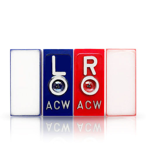
  •  Standard Mammography X-Ray Markers feature a standardized set of abbreviations created by the American College of Radiology (ACR). A good, quality set of mammogram x-ray markers will include R/L/M designations on separate markers and degree markers. The range of markers in these sets allows the technologist to properly identify and label specific mammography images with imprinting. Like the standard left/right markers, there is a standardized color scheme for easy identification of these specialized markers: red for right, blue for left, and yellow for medial.
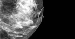
  • Mammography Skin Markers help technologists to provide clear and accurate visualization of nipples, moles, scars, palpable masses, and non-palpable areas of concern. The routine use of skin markers is considered a best practice for mammography as they help improve communication and reduce viewing time and the potential for retakes.

3D breast Markers

Types Of Skin Markers:

  • 3D Breast Tomosynthesis Markers  –  Clear visualization with the least potential for “slinky” artifacts in 3D mammography compared to other skin markers.
  • Mole Markers  –  Provide certainty that densities on mammography images are moles and not areas of concern.
  • Nipple Markers  –  Immediately identify nipple location as a landmark for precise measurement, help detect motion, and aid in positioning in profile.
  • Palpable Mass Markers  –  Readily identify symptomatic areas and provide permanent documentation that a palpable area was noted.
  • Point of Pain Area of Concern Markers  –  Area of concern or pain markers clarify the area marked by a square as different from any other topographic or palpable area of study during interpretation.
  • Scar Markers  –  Correlate architectural distortion from previous biopsy sites by delineating the exact location of incisions.

Magic X-Ray Markers is a Radiology Xray markers suppliers, founded in 2009, based in California offering exclusive collection of X-ray Markers and a vast assortment of top-notch Radiography Markers.

[email protected]| www.magicxraymarker.com

|Skin Markers |

X-Ray Markers: Lead vs. Lead-Free Characters


X-ray markers are used by radiologic technologists to indicate the left or right side of a patient’s body, as well as to provide other important information, such as the patient’s name, date of the exam, and anatomical markers.

Traditionally, X-ray markers have been made from lead, which is a dense metal that is easy to shape and provides good visibility on X-ray images. However, lead is a toxic material that can pose health risks to both patients and radiologic technologists who handle the markers. In addition, the disposal of lead X-ray markers can be a challenge, as they must be treated as hazardous waste.

Lead-free X-ray markers have become increasingly popular in recent years, as they provide a safer alternative to lead markers. These markers are typically made from materials such as aluminum or plastic, which are lightweight and easy to clean. However, They could be more expensive than lead markers but can be disposed of more easily.

Here are the pros and cons of each type:

Pros of Lead X-ray markers:

  • Lead is a dense material and is easily visible on an X-ray image, which makes it an ideal material for markers.
  • Lead X-ray markers are more durable and long-lasting than lead-free markers, and they can withstand high temperatures and rough handling.
  • Lead is a low-cost material, so lead X-ray markers are generally less expensive than lead-free markers.

Cons of Lead X-ray markers:

  • Lead is a toxic material, and prolonged exposure to lead can cause health problems. This is a concern for radiology technicians who handle X-ray markers frequently.
  • Lead X-ray markers are heavy, which can be a problem when they need to be attached to a patient’s clothing or body. The weight of the marker can cause discomfort and can shift during the procedure, which may affect the accuracy of the image.

Pros of Lead-free X-ray markers:

  • Lead-free X-ray markers are made from materials that are not toxic, so they are safer for radiology technicians to handle.
  • Lead-free X-ray markers are lightweight, which makes them more comfortable for patients to wear during X-ray procedures.
  • Some lead-free X-ray markers are made from eco-friendly materials, which can be beneficial for facilities that prioritize sustainability.

Cons of Lead-free X-ray markers:

  • Lead-free X-ray markers may not be as durable as lead markers and may need to be replaced more frequently.
  • Lead-free X-ray markers are often more expensive than lead markers.
  • Some lead-free markers may not be as visible on an X-ray image as lead markers, which can affect the accuracy of the image.
Overall, both lead and lead-free X-ray markers have their advantages and disadvantages, and the choice between them depends on individual preferences and institutional policies. Some healthcare facilities have opted to switch to lead-free markers to reduce their environmental impact and protect the health of their staff and patients, while others continue to use lead markers for their high visibility and durability.

Ultimately, the decision to use lead or lead-free X-ray markers will depend on the facility’s priorities and preferences. Both types of markers have their pros and cons, and it is up to the facility to decide which type best fits their needs.


Magic X-Ray Markers is a Radiology Xray Marker supplier, founded in 2009, based in California offering an exclusive collection of X-ray Markers and a vast assortment of top-notch Radiography Markers and Dosimeter Film Badges.

[email protected]www.magicxraymarker.com

Position Indicator X-Ray Markers That Don’t Need Tape

Position Indicator Markers

Self-Adhesive Position Markers

Position Indicator Markers on X-Ray images help Radiologists identify the Left and Right sides of the body part being examined, as well as the inclination or the angle between the patient in the erect and supine position. Many Radiology Technologists prefer X-Ray markers with beads because they easily can indicate a patient’s horizontal or vertical position and the direction of inclination on the images.

Position Indicators are most useful when the X-Ray exam must occur in an erect position. For instance, the abdominal film is routinely obtained with the patient in an erect position to reduce the risk for patients with suspected bowel obstruction, as well as to assess the air-fluid levels in the distended bowel. In these circumstances, the only way that a radiologist would be able to tell if the exam was performed upright is by using Position Indicator markers. Due to gravity, in an upright abdomen X-Ray exam, the beads will drop to the lower part of the ring, subsequently casting a shadow of the beads and the ring on the X-Ray film. Based on the shadow and using the ring as a reference point, Radiologists would be able to recognize the exam as an upright abdomen. However, in a supine or laying down position exam, the lead balls will sit in the center of the ring.

Traditionally the only way to add a position indicator marker onto an X-Ray cassette was by taping. Magic X-Ray markers are now for the first time offering Self-Adhesive Position Indicator markers which will never require any kind of adhesives or tape. These Self-Adhesive Position Indicator markers are washable, reusable, and in compliance with hospitals’ infection control and prevention policy. https://www.magicxraymarker.com/SELF-ADHESIVE-X-RAY-MARKERS/self-adhesive-position-indicator-markers.html

Founded in 2009 and based in Lake Elsinore, California, Magic X-Ray Marker started out as the manufacturer of the world’s one and only washable and reusable, self-adhesive X-Ray markers. Over the years we have listened to our customers and expanded our product lines to include a large variety of Aluminum, Plastic, Personalized, and Same-Day shipping markers, holders, and accessories. We also offer top-quality lead and lead-free characters, as well as molds, glitters, and epoxy so you can create your own X-Ray markers as you, please!


Self-Adhesive X-Ray Markers | Plastic X-ray Markers | Elite-Aluminum X-Ray Markers | Lead-Free X-ray Markers | Personalized & Hand-Crafted X-Ray Markers | Lead & Lead-Free Characters | DIY Package for X-Ray Markers |Skin Markers | X-Ray Marker Holders| Expert Support


Magic X-Ray Markers is a Radiology Xray Marker supplier, founded in 2009, based in California offering an exclusive collection of X-ray Markers and a vast assortment of top-notch Radiography Markers.

[email protected]www.magicxraymarker.com

Everything You Need To Know About X-Ray Markers

X-ray Markers, otherwise called: anatomical side markers, Pb markers, lead markers, x-ray lead markers, or radiographic film recognizable proof markers, are used to stamp x-ray movies, both in doctor’s facilities and in mechanical working environments, (for example, on plane parts and engines). They are used on radiographic pictures to decide anatomical side of body, date of the methodology, and may incorporate patient’s name.

Most X-ray markers with initials comprise of a privilege and a left letter with the radiographer’s initials. There are additionally accessible markers to show situating of the body e.g. prostrate, or as to time when performing systems, for example, an Intravenous pyelogram. The x ray Markers with initials are very useful in many ways. It has been proposed that radiographic markers are a potential fomite for tactless microscopic organisms, for example, MRSA, and that they ought to be cleaned all the time; this, be that as it may, is not generally done.

Some X-ray markers are made with toon pictures or sparkle included on the markers. This style marker, known as mapmakers, demonstrates right or left side and incorporates technologists’ initials. All the more essentially a cheap substance, Handi-Tak is used to hold these markers to the X-ray plate during imaging. This Tacky tape should be cleaned amongst uses and disposed of when obviously soiled.

When looking for x ray Markers with initials, it is important to look at all the aspects related to it. There are a lot of lead markers available today, and they’re not an entangled bit of hardware. All it truly is a lead letter encased in a material that is undetectable in an x-ray machine. In any case, these seemingly insignificant details are vital to numerous radiography divisions and focuses. 

At Magic X-Ray Marker, we provide a wide range of markers at the best rates. You can even customize it like the way you want. We are a hotspot for X-Ray Lead Markers including Aluminum and Plastic X-Ray Markers. We offer a substantial choice of sizes, shapes and colors including full units and in addition singular X-Ray Marker sets. Tweak your X-Ray Markers with your initials on generally things.

 Our lead markers are altogether made in-house utilizing quality plastic or potentially aluminum housings to guarantee life span and sublime quality. We can modify your markers to incorporate your company or division’s initials, and the greater part of our requests delivers within a few business days.

Radiation exposure increases risk of health problems for medical staff

Researchers found that health care workers who perform fluoroscopy for heart procedures are at greater risk for orthopedic problems, cataracts, skin lesions and cancers.
Study leader Maria Grazia Andreassi, PhD, head of the Genetics and Molecular Epidemiology Unit at the National Research Council Institute of Clinical Physiology in Pisa, Italy, and colleagues publish their findings in the journal Circulation: Cardiovascular Interventions.

Fluoroscopy is a form of medical imaging that uses a continuous X-ray beam to see real-time images of certain parts of the body.
The technique is used for a number of heart procedures, including coronary angiography – used to detect heart conditions – and coronary artery angioplasty – used to widen blocked or narrowed arteries.
Such procedures are normally conducted in a hospital’s cardiac catheterization (cath) lab.
Cardiologists, electrophysiologists have greatest radiation exposure

A number of medical staff in cath labs are exposed to fluoroscopy radiation, including doctors, nurses and technicians. Andreassi notes that, of all X-ray procedures, fluoroscopy-guided heart procedures lead to the greatest radiation exposure among health care workers.
“Interventional cardiologists and electrophysiologists have a two to three times higher annual exposure than that of radiologists, as they are closer to the radiological source and experience radiation exposure with the patient, whereas diagnostic radiologists are generally shielded from radiation exposure,” she explains.

Andreassi says that busy cardiologists and electrophysiologists are exposed to around 5 millisieverts (mSv) of radiation each year; mSv is a measure of how much radiation is absorbed by the human body.

This means that over a 30-year career, these health care workers may be exposed to around 50-200 mSv – the equivalent of 2,500-10,000 chest X-rays. But how does such exposure affect health? Andreassi and her team set out to investigate.